Introduction to Posterior Rhinoscopy
Posterior rhinoscopy is a classical yet highly valuable diagnostic technique used in the field of Otolaryngology (ear, nose, and throat medicine). While modern imaging and endoscopic tools have transformed clinical practice, posterior rhinoscopy still holds an important place, especially in settings where advanced equipment may not be readily available. At its core, the procedure allows clinicians to visualize the back of the nasal cavity and the upper part of the throat (nasopharynx) using a simple mirror and light source.
Unlike anterior rhinoscopy, which focuses on the front portion of the nasal cavity, gives access to deeper anatomical structures that are otherwise difficult to examine. This includes areas like the posterior nasal septum, choanae, and openings of the Eustachian tubes. These regions are critical when diagnosing certain conditions that cannot be detected through external examination alone.
Although it may seem like a basic technique, posterior rhinoscopy requires skill, patience, and a strong understanding of nasal anatomy. For medical students and practitioners alike, mastering this method offers not only diagnostic value but also a deeper appreciation for traditional clinical examination techniques that continue to remain relevant.
Historical Background and Evolution
Posterior rhinoscopy has a fascinating history that reflects the evolution of medical diagnostics. The technique dates back to the 19th century when physicians began experimenting with mirrors to visualize internal body structures. One of the pioneers in this area was Manuel García, who initially used mirrors to examine his own vocal cords, indirectly influencing ENT diagnostic methods.
As medical science progressed, posterior rhinoscopy became a standardized procedure in ENT practice. Physicians refined mirror design, improved lighting techniques, and developed better patient positioning methods. For decades, it served as one of the primary ways to inspect the nasopharynx before the invention of fiberoptic instruments.
In modern times, while nasal endoscopy has largely taken over due to its superior visualization, posterior rhinoscopy is still taught and practiced. It remains especially useful in resource-limited settings and serves as a foundational skill in ENT training programs worldwide. Its historical importance also highlights the ingenuity of early physicians who relied on simple tools to achieve complex diagnostic insights.
Anatomy Visualized in Posterior Rhinoscopy
Understanding the anatomy visible during is crucial for accurate diagnosis. The procedure primarily focuses on the nasopharynx, a region located behind the nasal cavity and above the soft palate. This area contains several key structures that play important roles in respiration and ear function.
Among the structures observed are the posterior nasal septum, the choanae (posterior openings of the nasal cavity), and the openings of the Eustachian tubes. The Eustachian tube openings are particularly important because they connect the middle ear to the nasopharynx and help regulate ear pressure. Any abnormality here can lead to ear-related symptoms such as hearing loss or fullness.
Additionally, clinicians can assess the adenoids, which are lymphoid tissues located in the nasopharynx. Enlargement of adenoids is common in children and can contribute to breathing difficulties, snoring, and recurrent infections. Posterior rhinoscopy provides a direct way to evaluate these structures without relying on imaging alone.
Instruments and Equipment Used
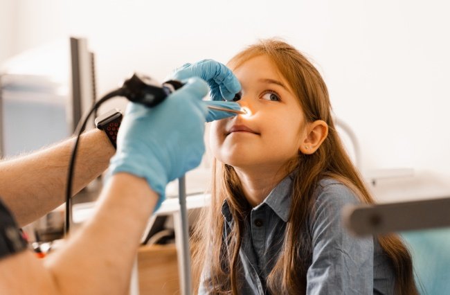
The primary instrument used in posterior rhinoscopy is the mirror. This small, angled mirror is attached to a handle and is inserted into the oral cavity to reflect the image of the nasopharynx. The mirror must be warmed before use to prevent fogging, which can obscure visibility.
A good light source is essential for the procedure. Traditionally, a head mirror or headlamp is used to direct light into the patient’s mouth and onto the mirror. Modern setups may include LED headlamps that provide brighter and more focused illumination, improving the quality of the examination.
Other supportive tools include a tongue depressor to keep the tongue out of the way and sometimes topical anesthetic sprays to reduce gag reflex. Although the equipment is simple compared to endoscopic systems, proper handling and coordination are key to obtaining a clear view.
Procedure: Step-by-Step Technique
Performing posterior rhinoscopy requires careful technique and patient cooperation. The patient is usually seated upright with their mouth open and tongue extended. A tongue depressor is used to hold the tongue down, allowing better access to the oropharynx.
The clinician then gently inserts the warmed mirror into the mouth, positioning it behind the soft palate. The mirror is angled upward to reflect the view of the nasopharynx. It is important to avoid touching the posterior pharyngeal wall, as this can trigger a gag reflex and make the procedure uncomfortable for the patient.
With practice, the examiner can quickly identify key structures and assess for abnormalities. The entire procedure typically takes only a few minutes but provides valuable diagnostic information. Proper communication and reassurance help ensure patient comfort and cooperation throughout the process.
Clinical Indications and Uses
Posterior rhinoscopy is used to evaluate a variety of nasal and nasopharyngeal conditions. One of its primary uses is in the assessment of adenoid hypertrophy, especially in children who present with nasal obstruction or mouth breathing.
It is also helpful in diagnosing infections, tumors, and structural abnormalities in the nasopharynx. For example, clinicians may use it to detect conditions like Nasopharyngeal carcinoma, which may not be visible through anterior examination alone.
Additionally, posterior rhinoscopy can aid in evaluating chronic conditions such as Sinusitis and Allergic rhinitis. By examining the posterior nasal cavity, clinicians can better understand the extent of inflammation or blockage and tailor treatment accordingly.
Advantages of Posterior Rhinoscopy
One of the biggest advantages of posterior rhinoscopy is its simplicity. The procedure does not require expensive equipment, making it accessible in many clinical settings, including rural and low-resource areas.
Another benefit is that it provides real-time visualization of the nasopharynx without radiation exposure. This makes it a safe option for repeated examinations, particularly in children or patients who require ongoing monitoring.
Moreover, it helps clinicians develop strong diagnostic skills. Because the technique relies heavily on anatomical knowledge and manual dexterity, it enhances a physician’s ability to interpret subtle findings that might otherwise be overlooked.
Limitations and Challenges
Despite its usefulness, has several limitations. One of the main challenges is patient discomfort, particularly due to the gag reflex. Some patients may not tolerate the procedure well, making it difficult to obtain a clear viewAnother limitation is restricted visibility. Compared to modern endoscopic techniques, posterior rhinoscopy offers a smaller and less detailed field of view. This can make it harder to detect subtle abnormalities or lesions.
Additionally, the procedure requires a certain level of expertise. Inexperienced practitioners may struggle with mirror positioning and interpretation of findings. As a result, it is often supplemented or replaced by nasal endoscopy in advanced clinical settings.
Comparison with Modern Techniques
In today’s medical practice, posterior rhinoscopy is often compared with nasal endoscopy. Endoscopy uses a flexible or rigid scope equipped with a कैमरा and light source, providing high-resolution images of the nasal cavity and nasopharynx.While endoscopy offers superior visualization, it also requires more advanced equipment and training. on the other hand, remains a quick and cost-effective alternative for initial assessment.Rather than being obsolete, complements modern techniques. It serves as a valuable screening tool and can guide the need for further investigation using endoscopy or imaging studies.
Training and Skill Development
Learning posterior rhinoscopy is an essential part of ENT training. Medical students and residents are taught the technique early in their education to build foundational examination skills.
Practice is key to mastering the procedure. Trainees must become familiar with mirror handling, patient positioning, and anatomical landmarks. Simulation models and supervised clinical practice can help improve proficiency.
Over time, clinicians develop the ability to perform the procedure quickly and accurately. This skill not only enhances diagnostic capability but also boosts confidence in clinical decision-making.
Patient Preparation and Comfort
Ensuring patient comfort is crucial for a successful posterior rhinoscopy. Before the procedure, the clinician should explain what will happen and address any concerns the patient may have.
Topical anesthetics can be used to reduce sensitivity in the throat and minimize gag reflex. Encouraging the patient to breathe slowly through the nose can also help relax the muscles and make the procedure easier.
A calm and reassuring approach goes posterior rhinoscopya long way in improving patient cooperation. When patients feel comfortable and informed, they are more likely to tolerate the procedure well.
Conclusion
Posterior rhinoscopy may seem like a traditional technique in an era dominated by advanced medical technology, but its relevance remains undeniable. It offers a simple, cost-effective, and efficient way to examine the nasopharynx and diagnose a wide range of conditions.